
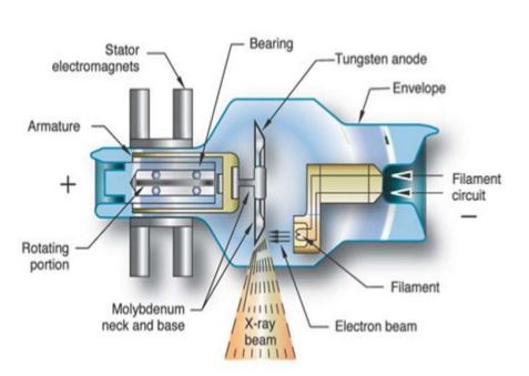
The x-ray tube provides an environment for the production bremsstrahlung and characteristic x-rays. It converts electrical energy into x-rays and heat. It is a relatively simple device containing only two principle elements: the cathode and the anode. Electrons are emitted from the cathode and then accelerated to the anode by a high electrical potential difference. They hit the andoe and lose energy by a series of reaction, producing X-rays and waste heat.
Electrons are released from the cathode filament by thermionic emission. The filament is made of tungsten as it must withstand strong ohmic heating. Most x-ray tubes are dual-filament design: the long one is used when large body parts are imaged while the short one is used when better spatial resolution is desired.
Because of the low efficiency, huge amount of heat is generated at the anode during operation. The temperature is so high that even tungsten, the strongest heat resistant metal, will melt quickly. Therefore, a rotating anode disc is used. The disc is made of tungsten insert embedded in a copper block. The copper is supporting the tungsten as well as the heat sink of the tungsten target. The anode is mounted on an induction motor and is spinning when the x-ray tube is operating. Electrons are striking a ring instead of a point and heat can be distributed on the anode uniformly.
X-ray tubes evolved from experimental Crookes tubes with which X-rays were first discovered on November 8, 1895, by the German physicist Wihelm Conrad Röntgen. The availability of this controllable source of X-rays created the field of radiography, the imaging of partly opaque objects with penetrating radiation. In contrast to other sources of ionizing radiation, X-rays are only produced as long as the X-ray tube is energized. X-ray tubes are also used in CT scanners, airport luggage scanners, X-ray crystallography, material and structure analysis, and for industrial inspection.


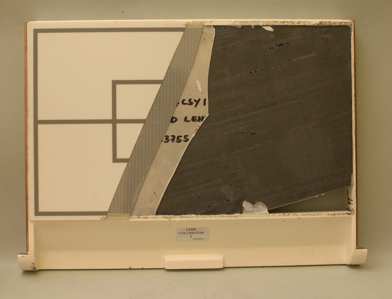
It is the collimator from rectilinear scanner used for restricting the detection to a small area directly below its position so that by the end of the scan emission from the whole study area has been detected.
A rectilinear scanner is an imaging device, used to capture emission from radiopharmaceuticals in nuclear medicine. The image is created by physically moving a radiation detector over the surface of a radioactive patient. The patient is administered with a radioactive pharmaceutical agent, such as iodine, which will naturally collect in the thyroid. The detector moves in a raster pattern over studied area of the patient, making a constant count rate. A collimator restricts detection to a small area directly below its position so that by the end of the scan emission from the whole study area has been detected. The output method is designed such that positional and detection information is maintained. For example when using a light source and film the light is moved in tandem with the detector, and the intensity of light produced increases with an increase in activity, producing dark areas on the film.
It has become obsolete in medical imaging, largely replaced by the gamma camera since the late 1960s.
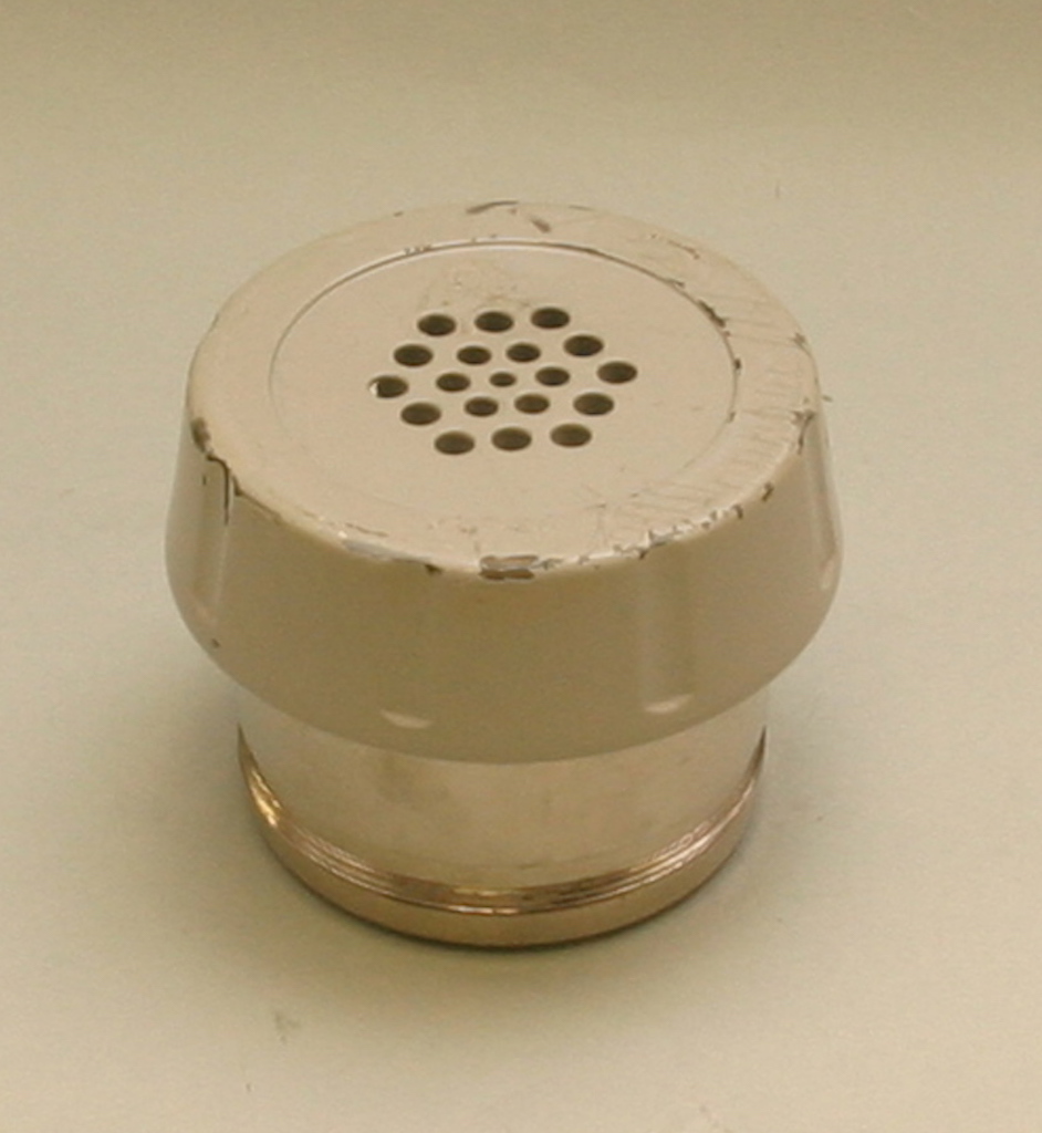
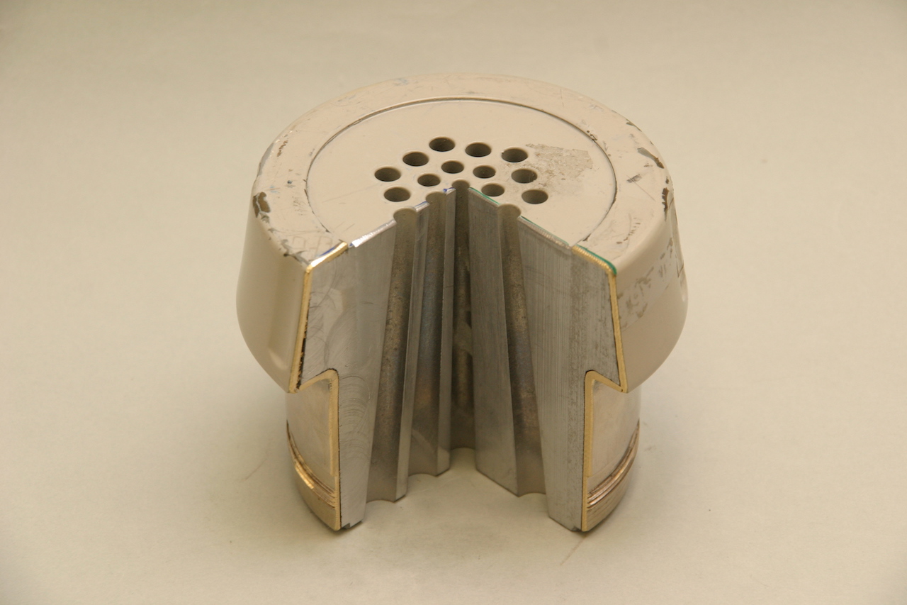
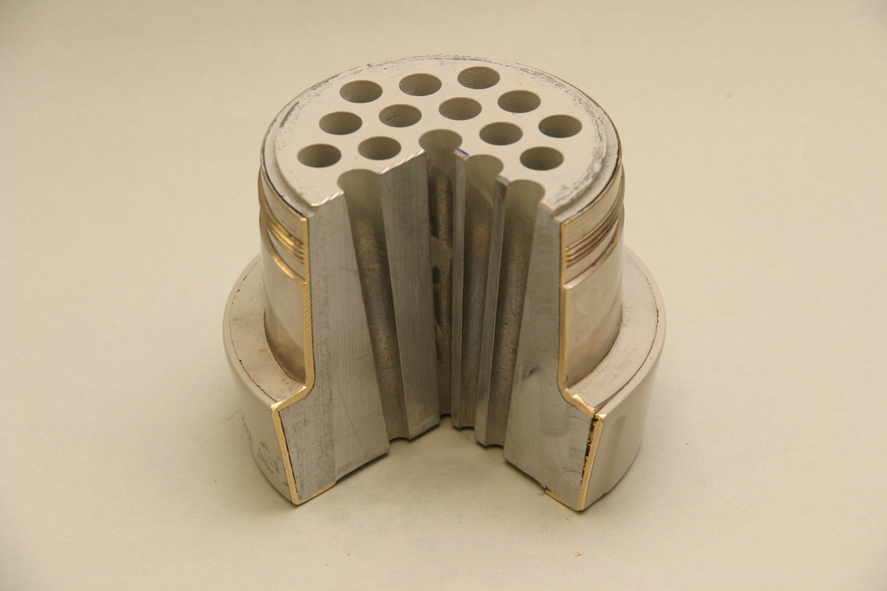
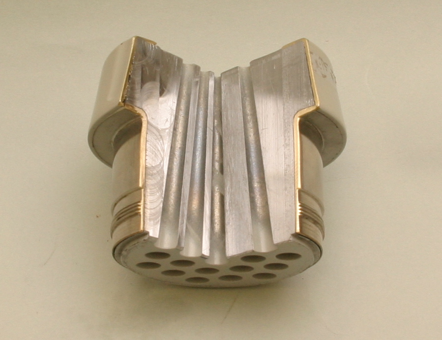
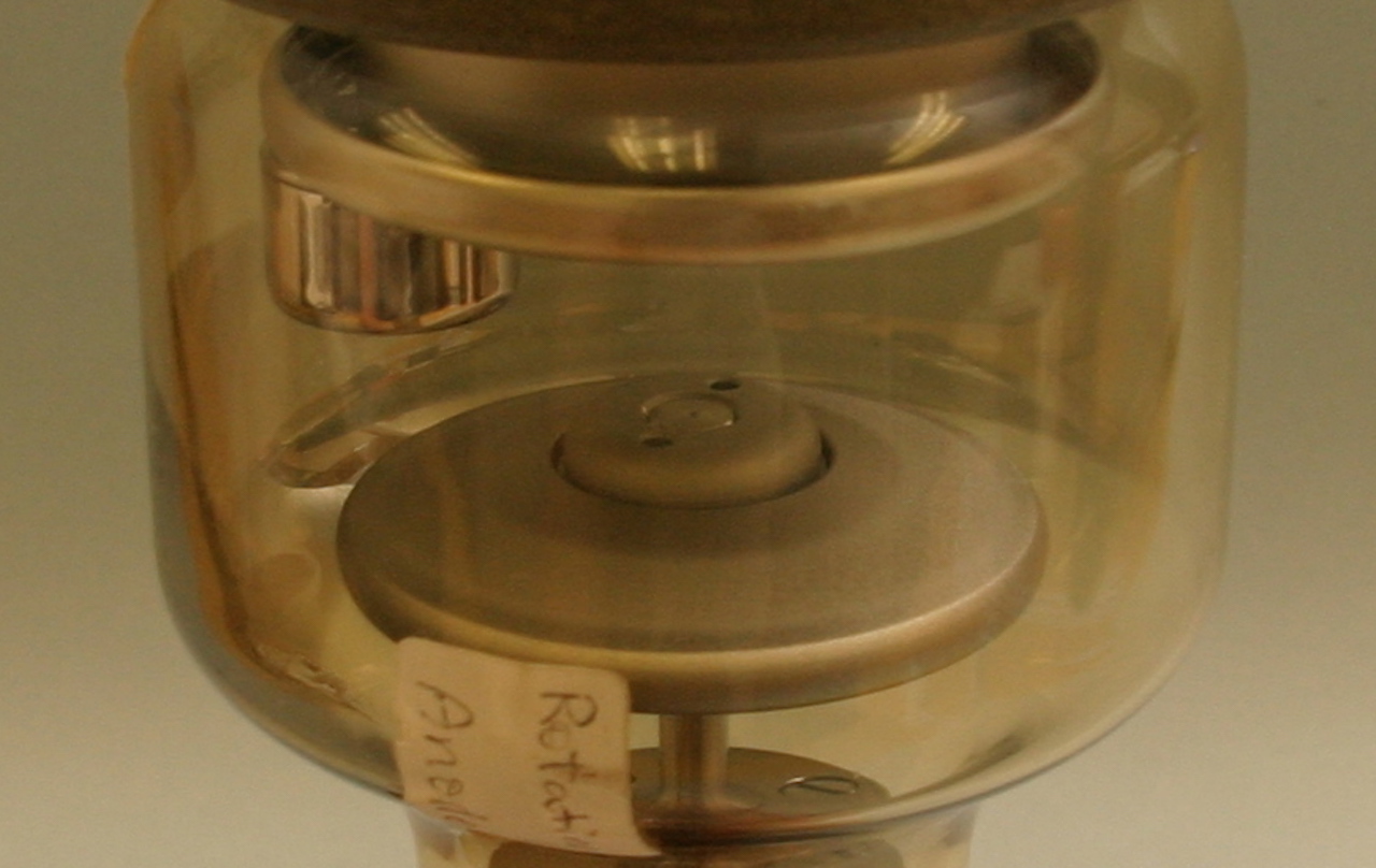
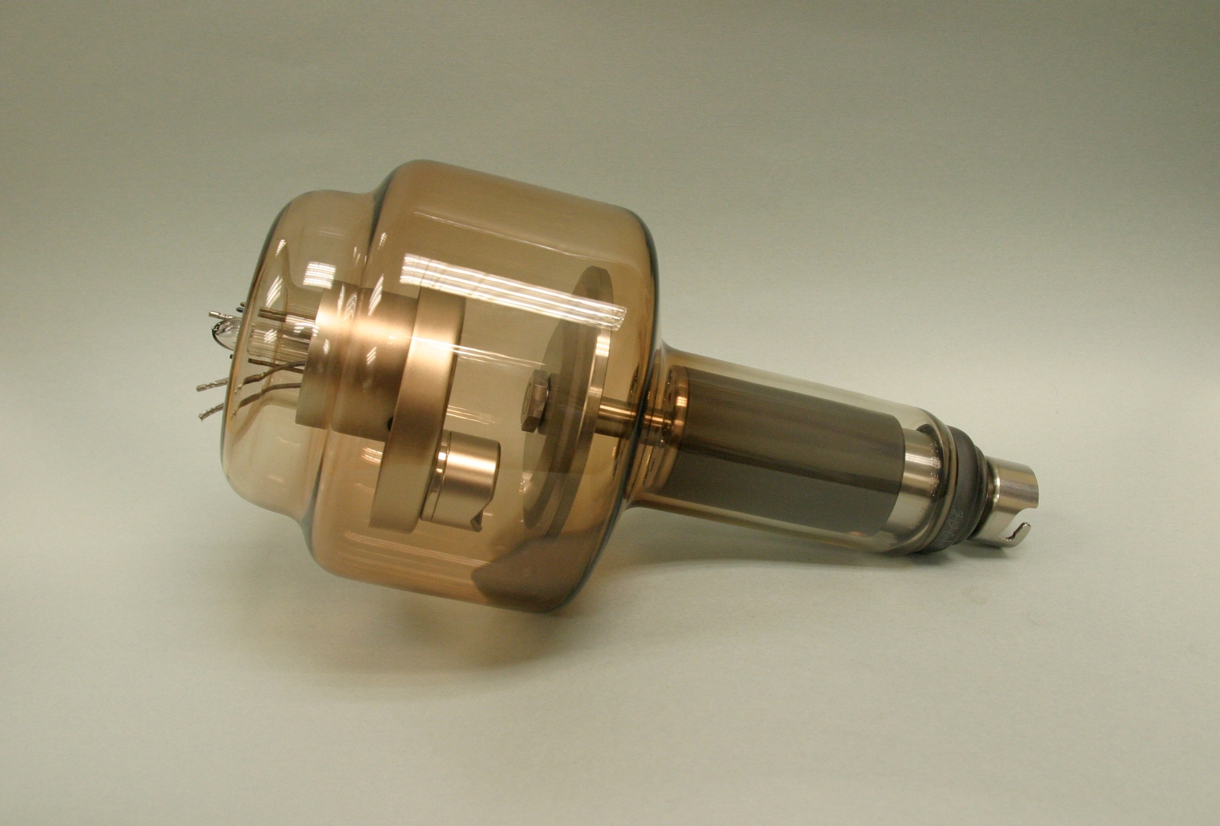
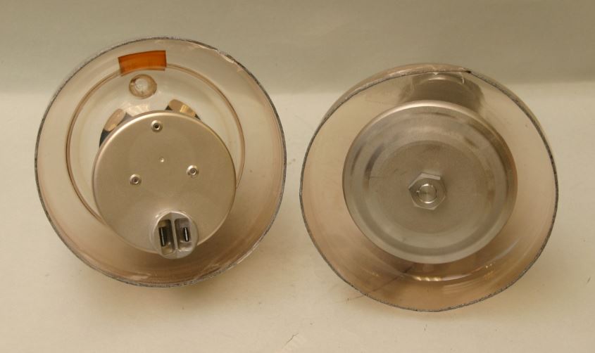
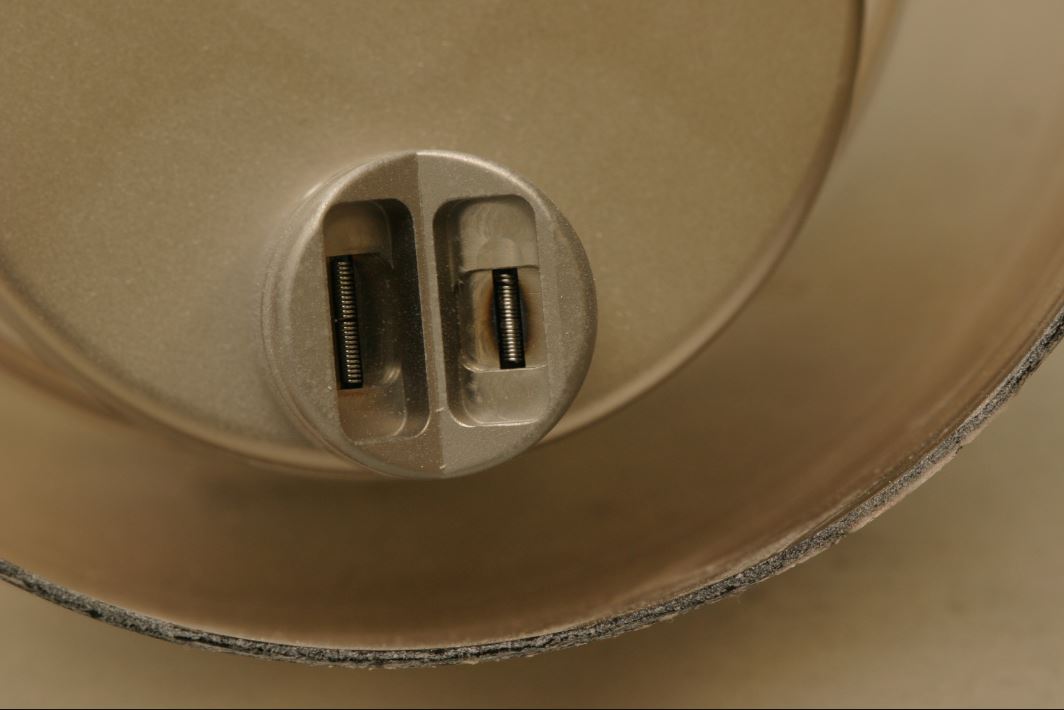
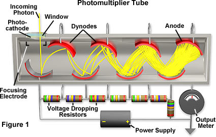
Photomultiplier (photomultipliers or PMTs for short), members of the class of vacuum tubes, and more specifically vacuum phototubes, are extremely sensitive detectors of light in the ultraviolet, visible, and near-infrared ranges of the electromagnetic spectrum. These detectors multiply the current produced by incident light by as much as 100 million times (i.e., 160 dB), in multiple dynode stages, enabling (for example) individual photons to be detected when the incident flux of light is low.
The combination of high gain, low noise, high frequency response or, equivalently, ultra-fast response, and large area of collection has maintained photomultipliers an essential place in low light level spectroscopy, confocal microscopy, Raman spectroscopy, fluorescence spectroscopy, nuclear and particle physics, astronomy, medical diagnostics including blood tests, medical imaging, motion picture film scanning (telecine), radar jamming, and high-end image scanners known as drum scanners. Elements of photomultiplier technology, when integrated differently, are the basis of night vision devices. Research that analyzes light scattering, such as the study of polymers in solution, often uses a laser and a PMT to collect the scattered light data.

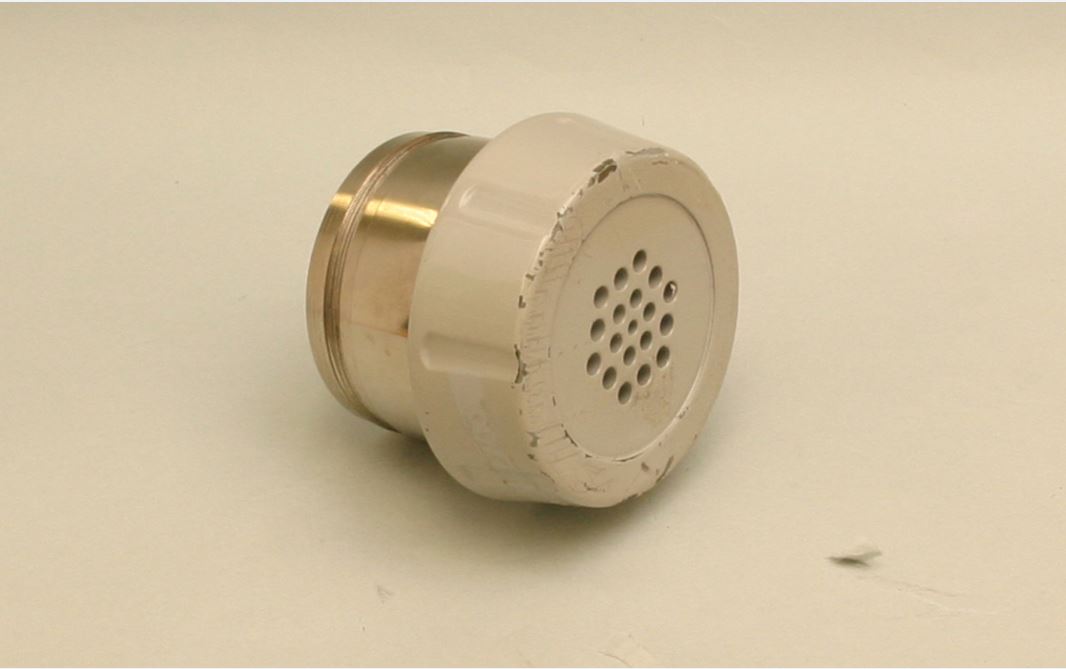
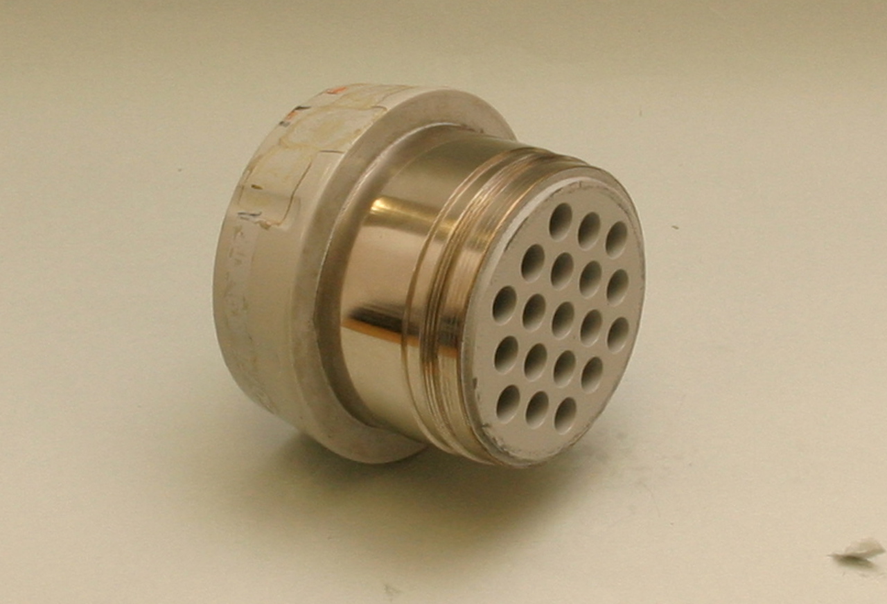
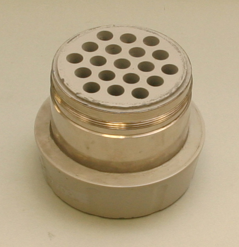
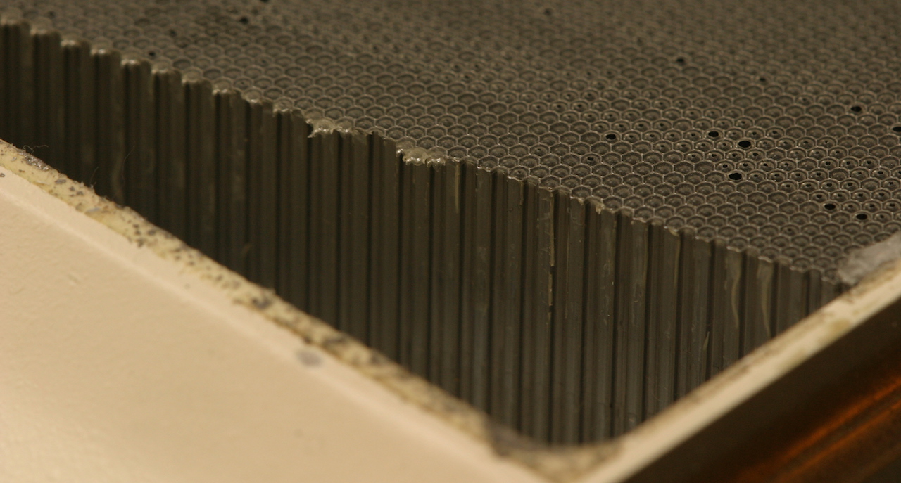
It is the Low Energy High-Resolution (LEHR) parallel hole collimator from single-photon Emission Computed Tomography (SPECT). SPECT is a nuclear medicine tomographic imaging modality. It is a powerful tool for quantifying the three-dimensional distribution of a radiotracer in humans, especially in oncology. It is considered to be the preferred modality in studying functional abnormalities between normal and cancer cells. In this process, the gamma rays from the administered radioisotope are encountered with photon attenuation and scattering. The gamma rays, before collision with the detector, encountered the collimator. The quality of the images in SPECT is seriously affected by the collimator's structure, properties of the detector, reconstruction algorithms, and also attenuation/scattering of gamma rays in the patient's body and the collimator. The collimation system is a critical component of a SPECT imaging system. Performance of a collimator was improved by increasing the absorption efficiency of the collimator material.
It is important to keep the septal penetration below 2%. Higher septal penetration will increase background noise, which can cause dramatic reduction in image resolution. In this collimator, all holes are parallel to each other. Low Energy High-Resolution (LEHR) collimators have high resolution images. They have a lot holes that are small and deep. The sensitivity is approx. 185,000 cpm for 1-uCi source, and the resolution is high with 0.65cm at 10cm from the patient side of the collimator.
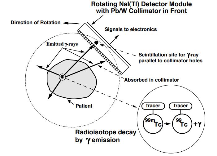
The SPECT devices usually have scintillation crystal (NaI scintillators). A 1 cm thick scintillator is perfectly adequate as it absorbs the majority of the incoming photons. Due to the relatively low gamma energy, a crystal with high light yield is needed, and NaI meets this requirement. In case of gamma cameras spatial resolution is limited primarily by the collimator (and because of the spatial blurring of the photomultipliers).
The NaI crystal is a hygroscopic material but fortunately this is not a considerable problem, since there are large contiguous crystals in SPECT devices (and in planar gamma cameras), which can be easily protected from humidity. However, due to the large size of the crystal it has to be mechanically resistant.