
Engineering Physics in Practice
The maintenance of sophisticated radiological equipment such as medical linear accelerator requires interdisciplinary knowledge and skills. Those who provide technical support to the equipment need a good grasp of electrical, electronic, mechanical, hydraulic, pneumatic microwave, vacuum, optics, safety engineering, general physics and medical physics principles in order to troubleshoot all kinds of faults as well as to safe guard the medical equipment operation.
1. Applied Physics to Linear Accelerator Maintenance
1.1 Beam Energy Control
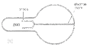
The basic physics of electron motion in the magnetic field is that the electron beam is deflected downwards as illustrated in Fig. 1a & 1b.
 |
 |
| Fig. 1. | (a) Electron beam travel in a straight line without the influence of magnet field. (b) Electron beam is bent in a curve path by the magnetic field. |
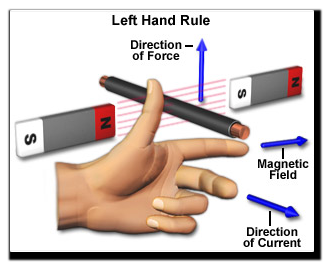
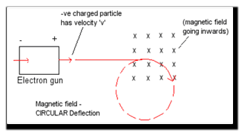
Fig. 2a explains Fleming's left hand rule that a force is exerted on an electron moving through the magnetic field. This force deflects the electron in a direction 90 degrees to the magnetic field which bends it in a circular motion as shown in Fig. 2b.
 |
 |
|
| Fig. 2. | (a) The left hand rule. | (b) Deflection of a moving electron in magnetic field. |
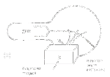
The bending magnets (Fig. 3) in a medical linear accelerator utilize magnetic deflection principle to focus an electron beam to hit the x-ray target or electron exit window in the treatment direction.
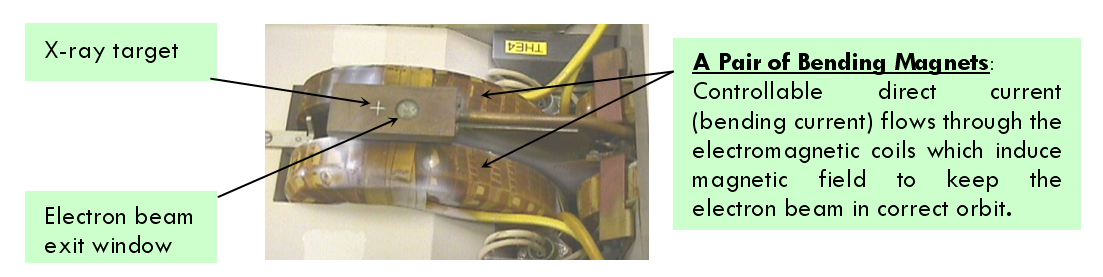 Fig. 3. Flight Tube and Bending Magnets of Elekta SL20 Linear Accelerator
Changing the bending current through the bending magnets (Fig. 4) will either increase or decrease the magnetic field strength which alters the electron beam path (Fig. 5 and 6) and hence its output beam energy (Fig. 7); a parameter that affects the clinical treatment.
 Fig. 4. Cross-section of bending magnet incorporating an accelerator guide shows the focusing effect by deflecting the beam in a correct path
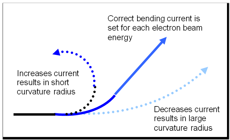
Fig. 5. The paths of electron beam affect by changing the bending current
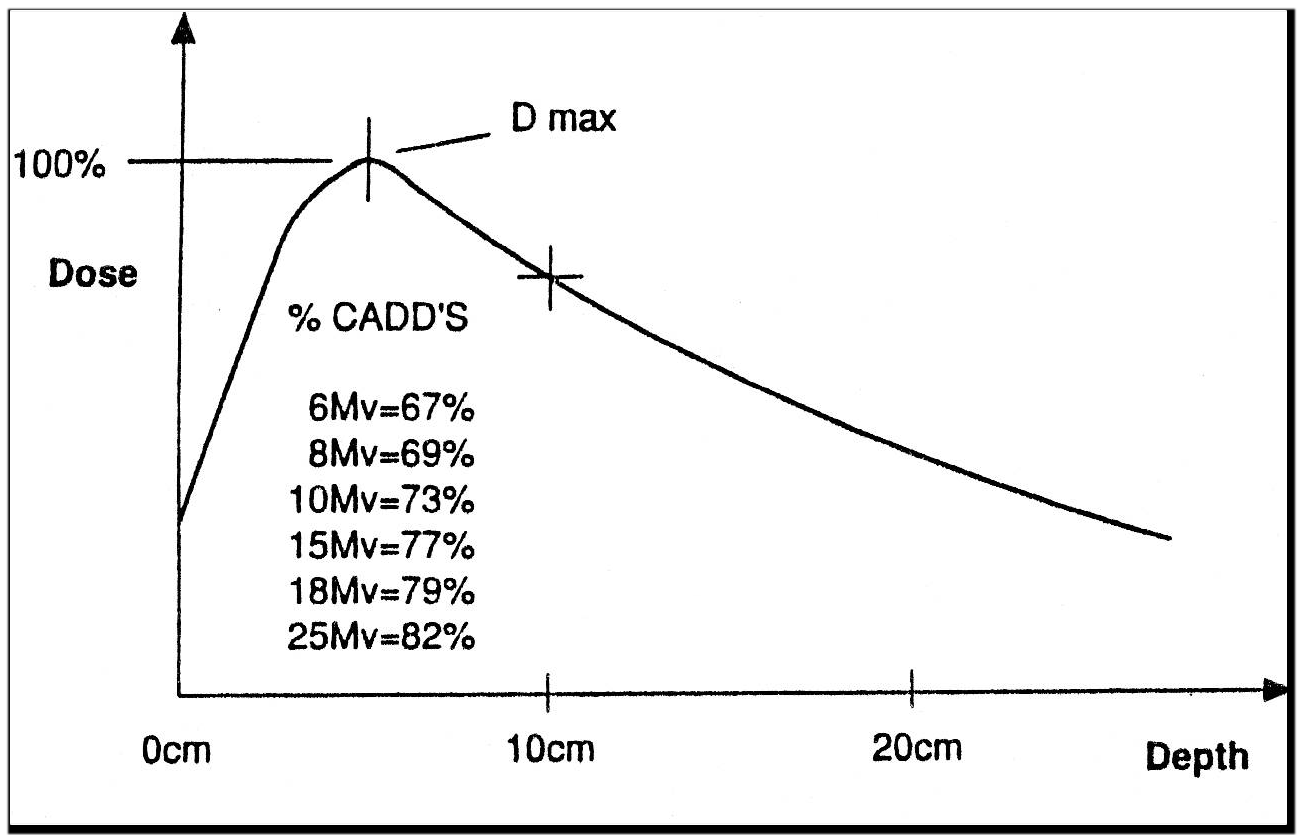
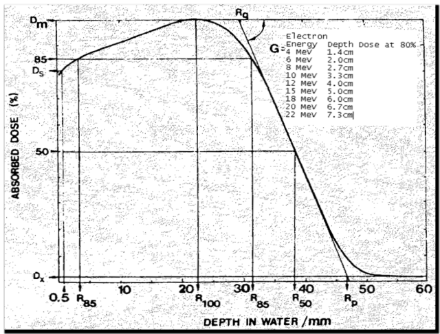
Medical linear accelerators producing either x-ray beam or electron beam is tuned to have a predetermined bending current for each beam energy. Any deviation of the preset bending current will alter the beam path (Fig. 6) and hence the output beam energy as measured in terms of depth dose curve (Fig. 7).
 |
| Fig. 6. The sladom type bending magnet of Elekta Linear Accelerator |
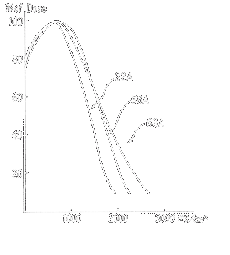 |
Fig. 7. Depth dose curve of 6 MeV electrons from linear accelerator. Variation of the bending current settings will affect the output beam energy. Bending current of 58A is the correct current for a 6MeV beam. Reducing the current to 22A causes a significant change in electron energy to 4MeV. Bending current of 68A shows a slight change in depth dose curve. |
|
Elekta linear accelerators employ 112.5° achromatic beam bending system which has three pairs of electromagnets as illustrated in Fig. 8 to keep the electron beam focussed and in the correct orbit.
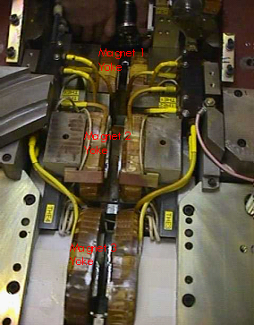 |
Fig. 8. Elekta Linear Accelerator employs 112.5° Achromatic Bending Magnets. Magnet 1 acts as an energy analyser by bending electron beam of the required energy to 45°. Magnet 2 reverses the bend bending to 45° to focus the beam in two orthogonal directions. Magnet 3 converges the beam to hit the x-ray target or electron exit window by bending it through 112.5°. |
As the x-rays and electrons beam energy are affected by bending magnet current, maintenance work such as bending magnets replacement or mechanical alignment; replacement or adjustment of bending magnets power supply; and electronic circuit boards replacement for controlling bending current should lead to verify the correct beam energy by depth dose measurement (Fig. 9).
 |
 |
| (a) Depth dose curve of x-ray beam | (b) Depth dose curve of electron beam |
Fig. 9. Depth dose measurements in water and field size 10 x 10 cm2.