
Engineering Physics Stream 工程物理科
History
It was the beginning of X-ray therapy service at Queen Mary Hospital in 1937 that led to the employment of physicists to maintain the radiation therapy equipment at the Queen Mary Hospital and the Queen Elizabeth Hospital in the 1960s. In the early eighties, physicists started to take up the role of acceptance testing and maintenance of diagnostic X-rays equipment. Nowadays, medical physicists (engineering physics) are working at 6 major Cluster Hospitals within Hospital Authority to provide maintenance support of radiological equipment.
 |
 |
 |
 |
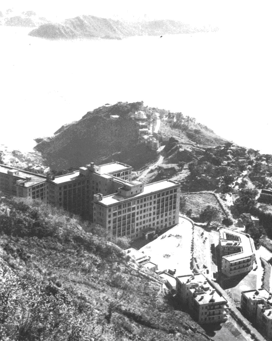
| Top left: | In 1937, X-ray therapy service was provided following the opening of Queen Mary Hospital which is situated at mid-level in Hong Kong Island, facing Lamma Island shown on top of the picture. (Photo Courtesy of Queen Mary Hospital) |
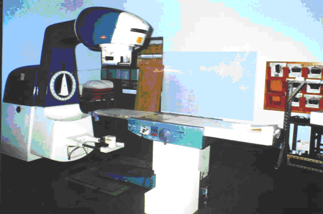
| Top right: | Commencement of Cobalt-60 teletherapy service in 1953 at the Queen Mary Hospital ended with the history of teletherapy in Hong Kong by decommissioning the AECL Cobalt-60 Teletherapy unit in 1993. |
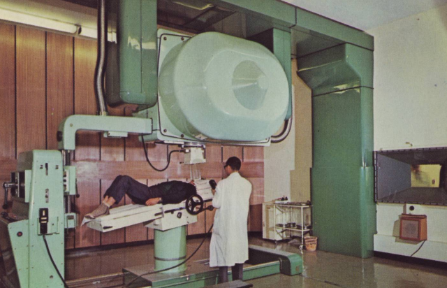
| Bottom left: | Brown Boveri Swiss-made Betatron at Queen Elizabeth Hospital in Kowloon operated at between 10 MeV and 35MeV. The patient was viewed from the control room through a 10 in. cube of high-density lead glass. |
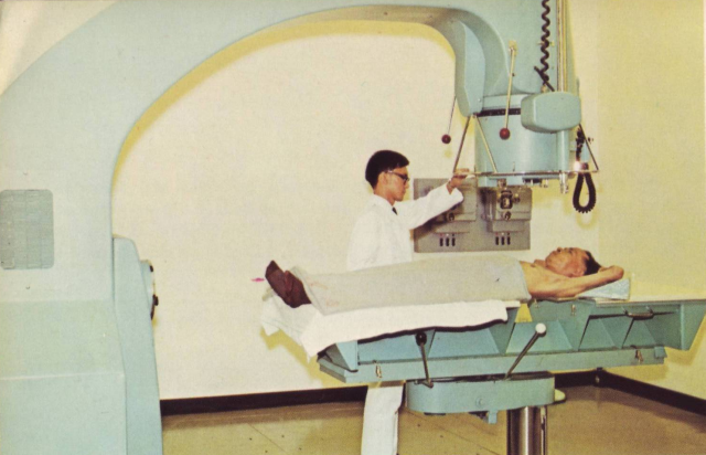
| Bottom right: | The installation of the first two 6MV medical linear accelerators by Vickers Armstrong (UK) at Queen Elizabeth Hospital in 1963. The first-generation linear accelerator powered by microwave tube to treat cancer. (Bottom Photos Courtesy of Public Works Department, Hong Kong) |
Medical Physicist (Engineering Physics)
Medical physicists (engineering physics) normally graduate with a University degree in electrical / electronic engineering, biomedical electronic, telecommunication or computer engineering. They are Chartered Engineers or professional engineers who are responsible for the management and technical support of radiological equipment. Certified Medical Physicists (Engineering Physics) are awarded to those experienced engineers who have passed the certification examination operated by HKAMP.
Engineering physics in the field of medical physics is the application of radiation physics and multidisciplinary engineering in support of radiotherapy and radio-diagnostic services within Hospital Authority hospitals in Hong Kong. Medical Physicists (engineering physics) should equip themselves with a wide range of engineering principles and have a strong background of physics to support engineering physics activities which are broadly identified into four core areas:
(i) Radiological Equipment Maintenance |
|
| Sophisticated radiotherapy equipment (e.g. medical linear accelerators, Integrated Brachytherapy equipment) and diagnostic x-rays equipment (e.g. CT scanners) are maintained by professorial engineers who lead the engineering team of X-Ray Workshop to perform mechanical, electrical and electronic maintenance of the radiological equipment. | 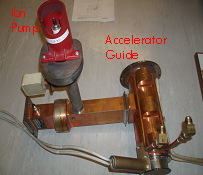 |
| The picture on the right shows the replacement of 6MV accelerator guide of Varian Clinac 6/100 due to short-circuit of ion pump. | |
(ii) Engineering Management |
|
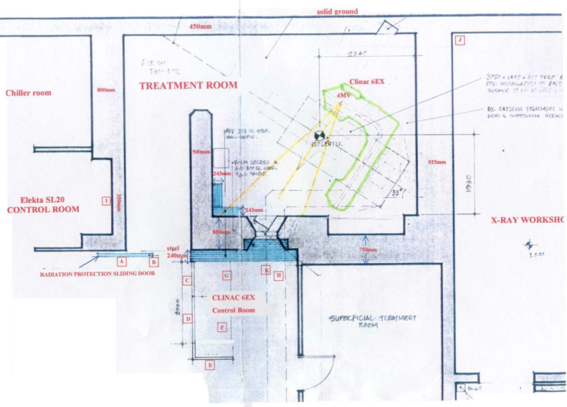 |
Engineering management is to lead engineering activities which include preventive maintenance inspection, quality assurance, medical electrical equipment safety, acceptance testing, and training. It is required to perform project management of equipment replacement / installation, equipment procurement, computer networking, new technology development, service contract, and legal-compliant for equipment operation. |
|
The picture on the left shows the site planning for the installation of linear accelerator. |
|
(iii) Radiological Equipment Quality Assurance |
|
| Dosimetric measurements, equipment calibration / alignment, patient dose audits and equipment commissioning are essential for the preservation of equipment performance standards. | 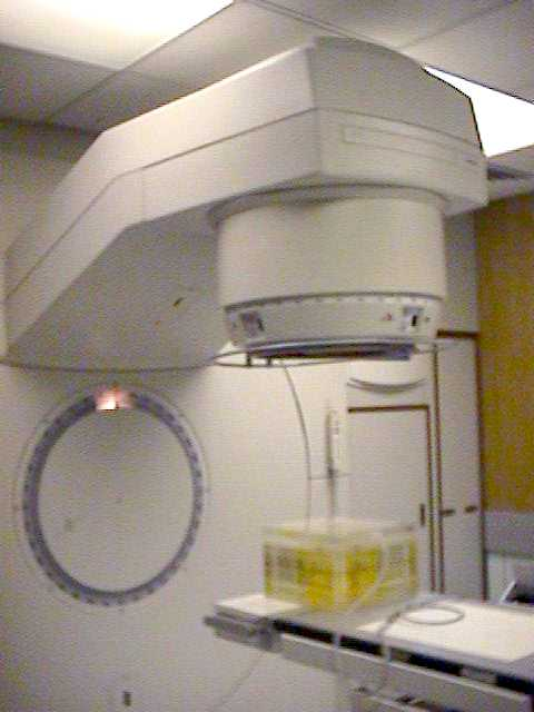 |
| The picture on the right shows the weekly dose output measurement of the linear accelerator as legally required by Radiation Ordinance of Hong Kong. | |
(iv) Radiation Safety |
|
 |
Radiation safety involves room design for radiaiton protection, measurements of leakage radiation, risk assessement and obligations of radiation protection supervisor. Career development has extended to non-ionization radiation safety involving medical equipment emitting laser and radiofrequency radiations. |
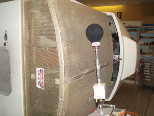
| The picture on the left shows the microwave leakage measurement from the magnetron which generates microwave to accelerate electrons inside the accelerator guide. | |
Engineering Physics in Practice
The maintenance of sophisticated radiological equipment such as medical linear accelerator requires interdisciplinary knowledge and skills. Those who provide technical support to the equipment need a good grasp of electrical, electronic, mechanical, hydraulic, pneumatic microwave, vacuum, optics, safety engineering, general physics and medical physics principles in order to troubleshoot all kinds of faults as well as to safe guard the medical equipment operation.
1. Applied Physics to Linear Accelerator Maintenance
1.1 Beam Energy Control
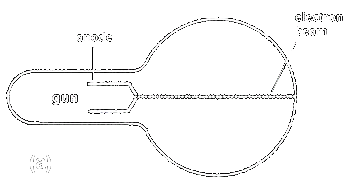
The basic physics of electron motion in the magnetic field is that the electron beam is deflected downwards as illustrated in Fig. 1a & 1b.
 |
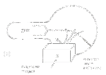 |
| Fig. 1. | (a) Electron beam travel in a straight line without the influence of magnet field. (b) Electron beam is bent in a curve path by the magnetic field. |
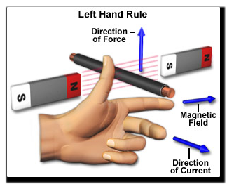
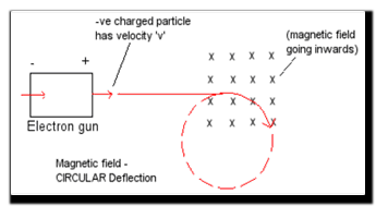
Fig. 2a explains Fleming's left hand rule that a force is exerted on an electron moving through the magnetic field. This force deflects the electron in a direction 90 degrees to the magnetic field which bends it in a circular motion as shown in Fig. 2b.
 |
 |
|
| Fig. 2. | (a) The left hand rule. | (b) Deflection of a moving electron in magnetic field. |
The bending magnets (Fig. 3) in a medical linear accelerator utilize magnetic deflection principle to focus an electron beam to hit the x-ray target or electron exit window in the treatment direction.
 Fig. 3. Flight Tube and Bending Magnets of Elekta SL20 Linear Accelerator
Changing the bending current through the bending magnets (Fig. 4) will either increase or decrease the magnetic field strength which alters the electron beam path (Fig. 5 and 6) and hence its output beam energy (Fig. 7); a parameter that affects the clinical treatment.
 Fig. 4. Cross-section of bending magnet incorporating an accelerator guide shows the focusing effect by deflecting the beam in a correct path
Fig. 5. The paths of electron beam affect by changing the bending current
Medical linear accelerators producing either x-ray beam or electron beam is tuned to have a predetermined bending current for each beam energy. Any deviation of the preset bending current will alter the beam path (Fig. 6) and hence the output beam energy as measured in terms of depth dose curve (Fig. 7).
 |
| Fig. 6. The sladom type bending magnet of Elekta Linear Accelerator |
 |
Fig. 7. Depth dose curve of 6 MeV electrons from linear accelerator. Variation of the bending current settings will affect the output beam energy. Bending current of 58A is the correct current for a 6MeV beam. Reducing the current to 22A causes a significant change in electron energy to 4MeV. Bending current of 68A shows a slight change in depth dose curve. |
|
Elekta linear accelerators employ 112.5° achromatic beam bending system which has three pairs of electromagnets as illustrated in Fig. 8 to keep the electron beam focussed and in the correct orbit.
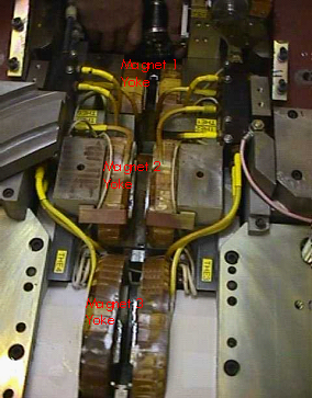 |
Fig. 8. Elekta Linear Accelerator employs 112.5° Achromatic Bending Magnets. Magnet 1 acts as an energy analyser by bending electron beam of the required energy to 45°. Magnet 2 reverses the bend bending to 45° to focus the beam in two orthogonal directions. Magnet 3 converges the beam to hit the x-ray target or electron exit window by bending it through 112.5°. |
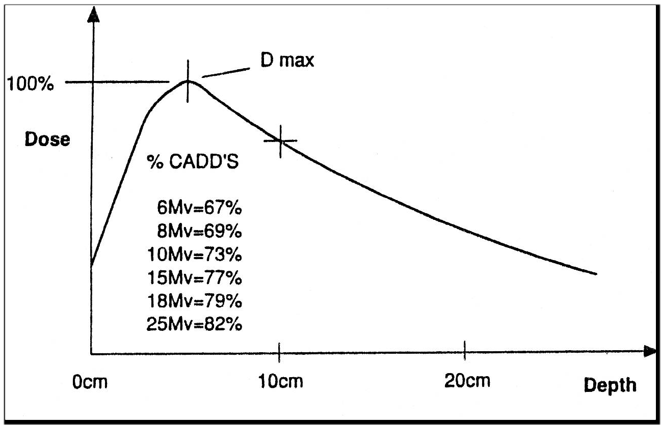
As the x-rays and electrons beam energy are affected by bending magnet current, maintenance work such as bending magnets replacement or mechanical alignment; replacement or adjustment of bending magnets power supply; and electronic circuit boards replacement for controlling bending current should lead to verify the correct beam energy by depth dose measurement (Fig. 9).
 |
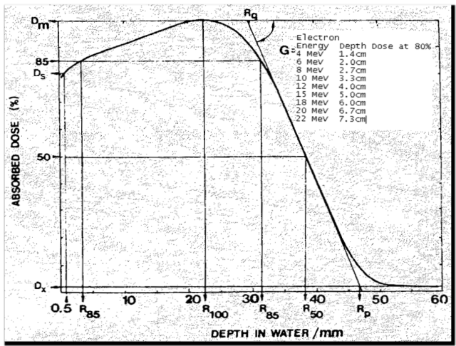 |
| (a) Depth dose curve of x-ray beam | (b) Depth dose curve of electron beam |
Fig. 9. Depth dose measurements in water and field size 10 x 10 cm2.
1.2 Practical Vacuum Physics
Vacuum plays an important part in linear accelerators (Linacs) and x-ray tubes which generate x-rays for medical and industrial applications. Why do linear accelerators need to operate at low gas pressure? Maintaining the vacuum in Linacs serves three purposes:
- To avoid oxidation of gun filament from burning out due to poor vacuum.
- To prevent collision of gas molecules with high velocity electrons, being accelerated across the accelerator guide.
- To inhibit microwave arcing which causes damage to accelerator structures.
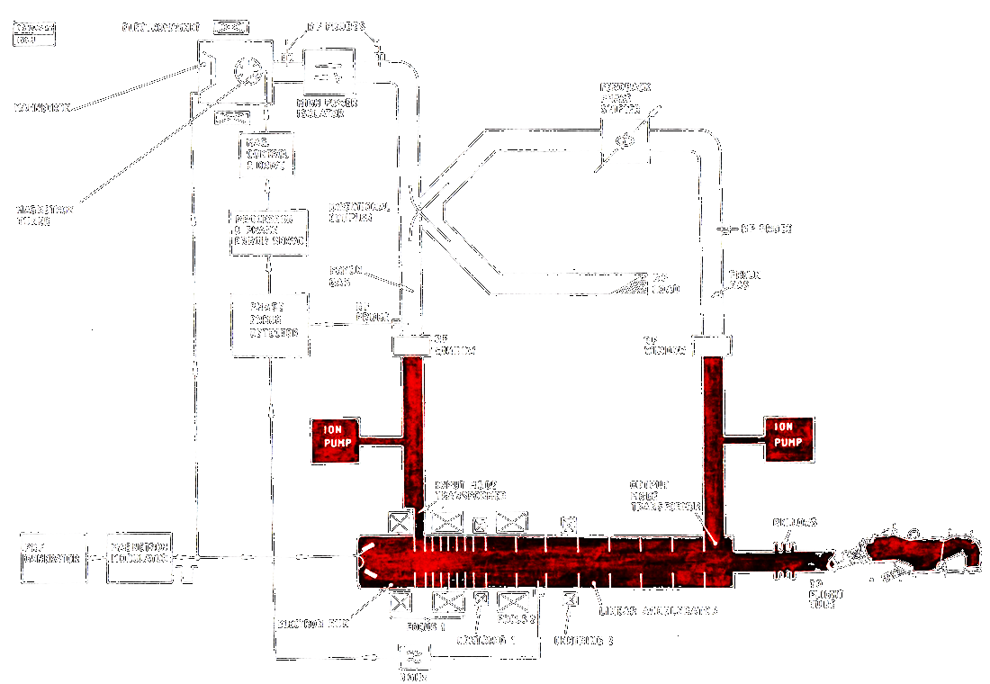 |
| Fig. 10. Schematic diagram shows components under vacuum (colour red) of Elekta Linac. |
The vacuum system of Elekta Linac (Fig. 10) includes electron gun, accelerator waveguide, flight tube (comprises of target / electron window assembly), ion pumps, input and output mode transformers. The key vacuum component is the ion pumps which serve to maintain high vacuum by gas ionization; and measure vacuum pressure by means of ion current within the entire vacuum system.
In case of vacuum breakdown due to outgassing (release of trapped gas atoms), vacuum leak or ion pumps fault, vacuum control interlock will stop Linac irradiation. Troubleshooting vacuum fault in Linac is not an easy task without the basic understanding of vacuum physics and techniques.
1.2.1 Basic Vacuum Physics
Gas molecules are in constant motion within the enclosed vessel (Fig. 11) such as the accelerator guide. Upon striking the vessel surface, they exert a force (F) on the wall of the vessel (A) where these molecular collisions result in a pressure rise. Pressure (P) in a vessel is referring to the impact force (Newton, N) on a unit area (square metre, cm2) caused by the gas molecules hitting the inner walls of the vessel.
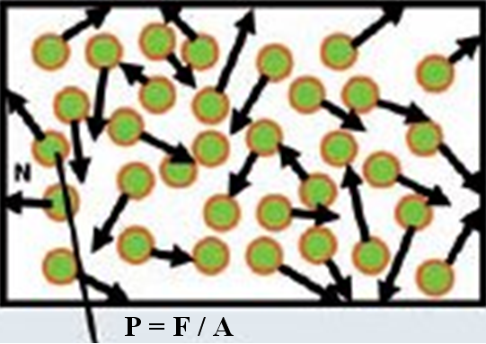
Fig. 11. Constant motion of gas molecules in a closed vessel
Vacuum is an empty space in which the pressure is significantly lower than atmospheric pressure. At atmospheric pressure, surfaces that are constantly bombarded by air and other gas molecules will quickly be contaminated (i.e. low or bad vacuum condition). By removing gas molecules until the pressure is reduced to a suitably low value (i.e. high or good vacuum condition), electrons can travel long distance without collisions with gas molecules in the accelerator guide. The process of removing gas molecules by ion pumps in the accelerator guide is known as evacuation.
To apply vacuum physics in troubleshooting vacuum fault, one should understand the ideal gas law, concept of pressure and vacuum relationship (Fig. 12).
 |
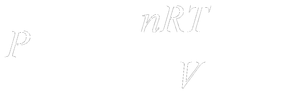 |
The pressure(P) in a vessel can be reduced by increasing the volume (V) of the vessel; decreasing the temperature (T) of the gas in the vessel; or by reducing the number of particles (n) in the vessel. R is the gas constant.
In practical situation, the ideal gas law can be applied to understand why the vacuum activity inside the accelerator guide is maintained.
- The accelerator guide is an enclosed volume under high vacuum condition (i.e. low gas pressure). Any pressure rise is caused by the increase of gas particles inside the guide and temperature of the guide.
- The working ion pumps will get hotter at its stainless steel surface under low vacuum condition. This is a good indication of high gas pressure (i.e. low vacuum) inside the accelerator guide which causes a temperature rise of ion pumps due to more gas molecules hitting its collecting electrodes.
The SI unit for pressure is the Pa (Pascal). The derived unit is the millibar (mbar). Another typical unit of pressure, millimeters of mercury (mmHg), is commonly used to show the atmospheric pressure 760 mm of Hg at sea level. In vacuum work, Torr is in common use as an indication of vacuum condition in linear accelerators. The conversion between different pressure units is 1 Torr = 1mm Hg = 1.333 mbar = 133.3 Pa. A good vacuum in accelerator guide is normally maintained at about 10-7 - 10-8 Torr.
Degree of vacuum which is the measurement of gas pressure can be divided into four pressure ranges:
| Low vacuum | 103 - 100 mbar | 750 - 0.75 Torr |
| Medium vacuum | 100 - 10-3mbar | 0.75 - 7.5-4Torr |
| High vacuum | 10-3 - 10-7mbar | 7.5-3 - 7.5-8Torr |
| Ultra-high vacuum | 10-7 - 10-12mbar | 7.5-8 - 7.5-13Torr |
 |
| Fig. 12. Diagram shows the relationship of gas pressure and vacuum level. |
Sorry! The followings are still under construction
1.2.2 Sputter Ion Pump
1.2.3 Vacuum Pressure Measurement
1.2.4 Vacuum Leak
1.3 High Frequency Power Transmission
2. Applied Physics to Diagnostic X-rays
2.1 Tube Current Fault
2.2 Incorrect kV measurement
2.3 Thermionic Emission
3. Applied Physics to Radiation Safety
3.1 Ionization Radiation Safety
3.2 Non-Ionization Radiation Safety